Human Distal Lung Organoid showing club cells and ciliated cells, everted for 10 days. Acquired with ZEISS LSM 900, Plan-Apochromat 63× / 1.40 Oil DIC M27. Courtesy of Prof. C. Kuo, Department of Medicine, Hematology Division, Stanford University, USA.
Courtesy of Prof. C. Kuo, Department of Medicine, Hematology Division, Stanford University, USA.

Empowering Research at Victoria House

with ZEISS and the Pioneer Group

Welcome to the Innovation Suite at Victoria House, where cutting-edge research meets world-class technology. Through a strategic partnership between ZEISS, a global leader in optical and optoelectronic technology, and Pioneer Group, we are thrilled to offer tenant researchers unparalleled access to advanced imaging solutions. Located in the heart of London, Victoria House is a hub for collaboration among life sciences businesses, technology entrepreneurs, academia, and large pharma teams.

Our partnership is designed to empower researchers with state-of-the-art microscopy tools, fostering a collaborative ecosystem that accelerates breakthroughs in drug discovery, cell biology, and biomedical research.  

  • Access to Advanced Imaging Technology
  • Comprehensive Support and Training
  • Microscopy Troubleshooting

Partnership

In Collaboration With
  • The Pioneer Group Logo

    The Pioneer Group

    The Pioneer Group supports science and life sciences businesses by providing specialised lab and office spaces across the UK. They offer incubation, mentoring, and access to funding to help startups grow. Their focus is to create collaborative ecosystems that accelerate innovation in biotech, medtech, and healthtech.

  • Victoria House London Logo

    Victoria House London

    Victoria House London offers 300,000 sq ft of modern lab, office, and amenity space. Home to leading life sciences organisations—from startups to global pharma—it supports growth with flexible lab rentals and business services. Pioneer Group provides on-site support, networking, and venture-building programs.

The front of Victoria House Bloomsbury London

Victoria House Bloomsbury London

Empowering Research Through Partnership

What the ZEISS partnership means for you — gain priority access to advanced imaging systems, expert guidance, and a platform to push the boundaries of your research.

  • Access to Advanced Imaging Technology: ZEISS provides tenant researchers with the Celldiscoverer 7 with LSM900, a high-performance confocal microscope, and the Axiovert 5 digital imaging system, both equipped with AI-driven tools to enhance research capabilities. 
  • Collaborative Ecosystem: Victoria House facilitates collaboration among startups, research groups, and industry experts, enabling tenants to leverage ZEISS expertise and technology for groundbreaking innovations.
  • Comprehensive Support and Training: The ZEISS team offers training and support to resident scientists, maximizing the impact of the technology and facilitating collaborative research opportunities.
  • Innovation Suite Amenities: Located on the seventh floor, The Incubator provides a 'one-stop shop' for startups and growing businesses, offering state-of-the-art equipment, shared services, and pre-negotiated discounts on consumables.
  • Engagement Opportunities: Participate in 'lunch and learn' events, talks from key opinion leaders, and on-site pop-up imaging clinics, all designed to showcase application topics and foster networking.

ZEISS Imaging Solutions at Victoria House

Reserve time to accelerate your research and discover powerful microscopy solutions.
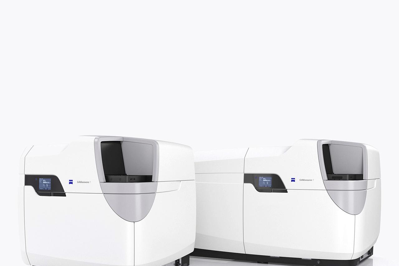
  • ZEISS Celldiscoverer 7

  • ZEISS Axiovert 5 Digital

    ZEISS Axiovert 5 Digital

  • ZEISS arivis Software

  • A small and large ZEISS Celldiscoverer 7 system

    ZEISS Celldiscoverer 7

    ZEISS Celldiscoverer 7

    with LSM900

    The ZEISS Celldiscoverer 7 with LSM 900 is a cutting-edge, fully integrated confocal imaging platform designed to accelerate biomedical research.

    This high-performance system delivers high-speed, high-resolution imaging with minimal user intervention—ideal for live-cell imaging, 3D reconstruction, and complex experimental workflows. Automatic calibration and intelligent focus tracking ensure optimal imaging conditions and reproducible results across experiments.

    Highly adaptable and user-friendly, the Celldiscoverer 7 offers a range of imaging modes and is optimized for flexibility, giving you the freedom to tailor the system to your specific research needs.

    Book Your Session
    The Celldiscoverer 7 is available now in the Innovation Suite. Visit the suite to learn more and book time on the system.

    Explore More
    Want to dive deeper into the automation and imaging features of the Celldiscoverer 7? Check out these curated resources.

  • ZEISS Axiovert 5 Digital

    ZEISS Axiovert 5 Digital

    ZEISS Axiovert 5 Digital

    Smart widefield microscopy at your fingertips

    The ZEISS Axiovert 5 Digital is a next-generation inverted microscope designed for efficient and intuitive imaging—no additional computer required.

    This all-in-one digital microscope combines high-quality optics with an integrated camera and smart documentation features. With just one push of a button, you can capture crisp images, add scale bars, and export data directly to your preferred storage—streamlining your workflow from image acquisition to analysis.

    Engineered for cell culture, routine imaging, and training applications, the Axiovert 5 Digital supports brightfield, phase contrast, and fluorescence imaging. It’s an ideal solution for researchers looking for quick, reliable, and easy-to-use microscopy without compromising on quality.

    Book Your Session
    The Axiovert 5 Digital is available in the Innovation Suite. Stop by to explore its capabilities and book your session.

    Explore More
    Want to make the most of the Axiovert 5 Digital and its AI capabilities? Check out these helpful resources.

  • ZEISS arivis software on a computer screen

    ZEISS arivis

    AI-Powered Image Analysis – Coming Soon to Victoria House

    The ZEISS arivis Advanced Image Analysis Ecosystem is a powerful suite of software tools designed to help researchers extract meaningful insights from complex microscopy datasets—quickly, accurately, and at scale.

    As microscopy data grows in volume and complexity, researchers often face bottlenecks in analysis, storage, and computing. The arivis platform addresses these challenges with AI-assisted workflows, support for multiple file formats, and optimized performance for large, multi-dimensional datasets. It integrates seamlessly with a wide range of microscopes—including the Celldiscoverer 7, available in the Innovation Suite at Victoria House.

    Whether you’re just starting with image analysis or looking to automate your pipeline, we can work with you directly to tailor your workflow, from data handling to AI model integration—adapting to your specific research needs and preferred level of automation.

    With no coding required and an intuitive interface, arivis empowers researchers to:

    • Automate and scale complex image analysis
    • Train and apply AI models within your workflow
    • Support diverse image formats and acquisition systems

    Coming Soon to the Innovation Suite
    ZEISS arivis will soon be available for researchers at Victoria House. Connect with our team to learn more, explore use cases, or request support in building your custom imaging pipeline.

    Explore More
    Want to make the most of the ZEISS arivis and its AI capabilities? Check out these helpful resources.

Be First to Know – ZEISS News & Tools for Researchers!

Pharma and Biotech Newsletter

Be the first to hear about ZEISS innovations, software updates, expert content and real life case studies—straight to your inbox.

Research in Action: Case Studies and Applications of Featured Imaging Technologies

Dive into real-world case studies and webinars that highlight how AI-enhanced imaging technologies are helping researchers model disease, analyze complex tissues, and bring safer, more effective drugs to market.
  • Understanding and Predicting Drug-Induced Liver Damage - hepatocyte hypertrophy is a major cause of drug failure in clinical trials. Researchers like Simon Plummer are using AI-driven imaging to better model liver responses and improve drug safety.

  • AI-driven imaging is unlocking the full potential of complex in vitro models (CIVM), overcoming the limits of 2D analysis for 3D/4D data. This shift—accelerated by FDA recognition—enables scalable, automated, and biologically relevant drug discovery using advanced microscopy and analysis tools.

  • Watch a video demo of ZEISS arivis Pro using machine learning to segment organoid structures and study Wnt signaling in maturation. Once trained, the analysis runs at scale via parallel processing in ZEISS arivis Hub for high-content 3D analysis.

  • Learn how to study Wnt signaling in intestinal organoids using simple imaging and AI-powered analysis. Explore how machine learning enables segmentation of organoid layers, nuclei, and cell bodies for deeper biological insights.

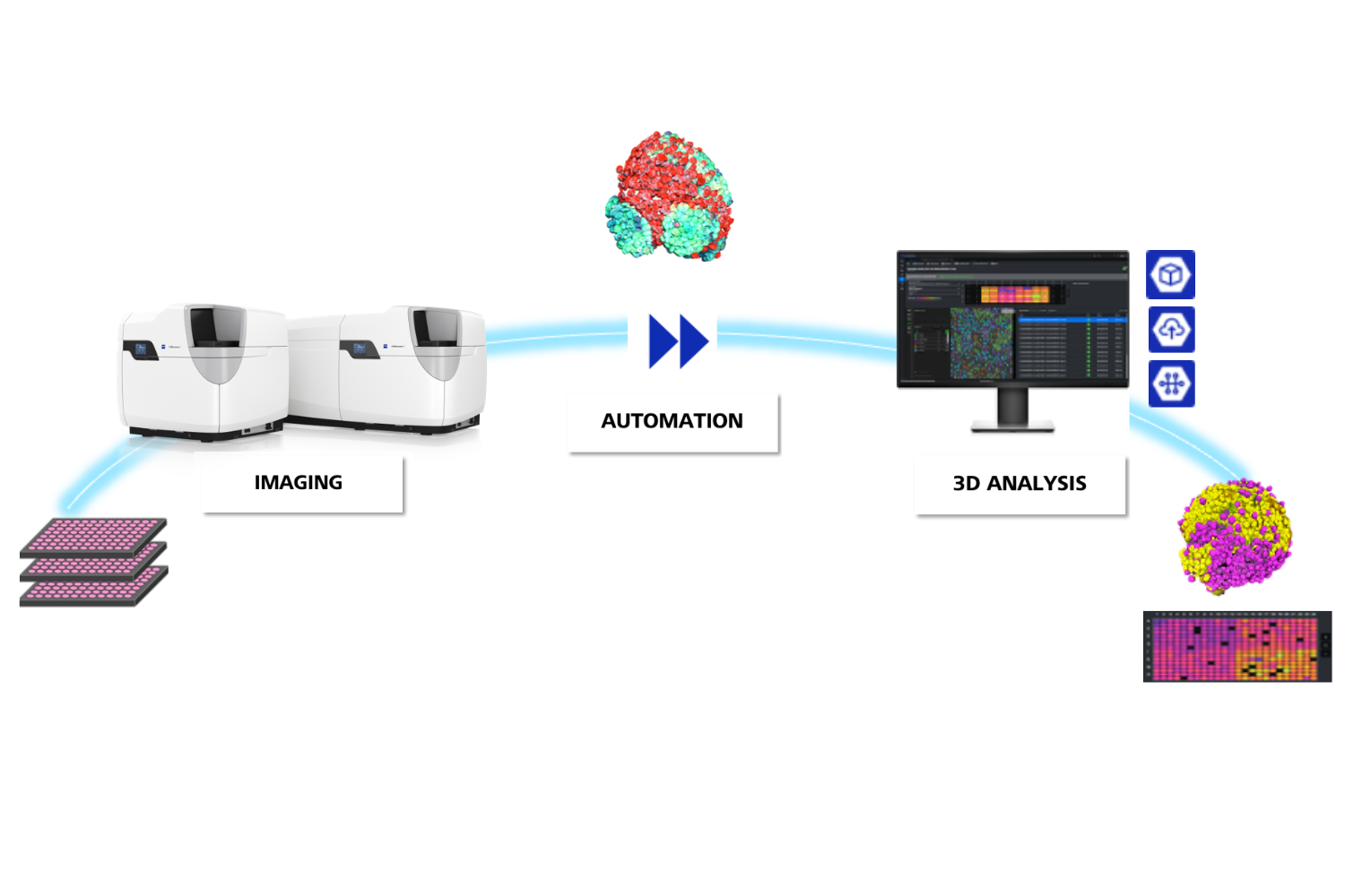
  • Evaluate drug-induced cytotoxicity more efficiently with an AI-powered workflow combining widefield imaging, HCS, and automated analysis on the ZEISS Celldiscoverer 7. This approach enables precise cell segmentation, viability assessment, and faster identification of effective treatment strategies in biopharma research.

  • Traditional 2D cultures fall short in predicting drug responses, but advanced Organ-on-a-Chip models paired with high-resolution 3D imaging are changing the game.

    Using the ZEISS LSM 910 with Airyscan, researchers can visualize detailed cellular behavior and gene expression, enabling earlier toxicity detection and more confident drug development.

  • Explore how 3D High Content Analysis and AI-powered imaging workflows are transforming biotech and pharma research. Learn how our solutions enable scalable, high-throughput 3D imaging with accurate segmentation and reproducible insights—accelerating discovery and reducing variability.

Meet the Team

Our dedicated ZEISS specialists are on hand to answer any questions you have about this partnership and to provide technical support for our imaging solutions in the innovations suite.
Dr. Emily Spencer
Dr. Emily Spencer Business Development Manager - Biotech & Pharma
Mr. Simon Denham
Mr. Simon Denham Account Manager – South Region
Dr. Nathan Cook UK Product & Application Sales Specialist
Dr. Emily Spencer
Dr. Emily Spencer Business Development Manager - Biotech & Pharma
Mr. Simon Denham
Mr. Simon Denham Account Manager – South Region
Dr. Nathan Cook UK Product & Application Sales Specialist

Upcoming Events

Stay tuned for information on seminars, networking opportunities, training sessions, and 'lunch and learn' events - coming soon to Victoria House.

Additional Resources

Contact Us

Click to complete a short form to get in touch.

Form is loading...