 and high-density minerals titanomagnetite and ilmenite (yellow) in chips of dolerite within concrete")
 and high-density minerals titanomagnetite and ilmenite (yellow) in chips of dolerite within concrete")
Advanced Solutions for Engineering Materials
Enhance Your EPSRC Strategic Infrastructure Funding ApplicationUnlock funding opportunities with cutting-edge microscopy. Let’s discuss how ZEISS can support your application.
Unlocking the full potential of current and future engineering materials starts with a deep understanding of their microstructure. Our microscopy solutions enable comprehensive analysis by combining morphological, chemical, and physical property studies, providing a complete picture of material behavior at the microscopic level. Read on to learn more about our solutions and contact us to explore how we can support your application.
Case Studies: Advancing Research with ZEISS Microscopy
Discover how our cutting-edge microscopy solutions are driving innovation in engineering materials research.. Internal tomography of targeted ROI scan shown in bright colours in A and B. Using DeepScout improves the contrast between different constituents, the visibility and identification of small grains within the matrix, and the outline and features within high-density grains (C and D).")
. Internal tomography of targeted ROI scan shown in bright colours in A and B. Using DeepScout improves the contrast between different constituents, the visibility and identification of small grains within the matrix, and the outline and features within high-density grains (C and D).")
Analysis of Concretes and Cements Using AI-enhanced X-ray Microscopy
The Global Cement and Concrete Association (GCCA) reports that over 14 billion cubic meters of concrete and more than 4 billion tonnes of cement are produced annually, highlighting the need for detailed study of these critical load-bearing materials to ensure optimal performance and safety. X-ray microscopy (XRM), a non-destructive 3D imaging technique, is transforming the analysis of concrete and cement by enabling the classification of grains, particles, interfaces, and pores at various scales, thus enhancing the understanding of their physical and compositional properties. A significant focus in XRM is on improving data acquisition and processing for accurate segmentation, which is crucial for quantifying different minerals or phases within these materials.
Advances in reconstruction techniques, particularly those utilizing AI algorithms, are addressing challenges in segmenting complex cement and concrete microstructures by reducing artefacts and noise, improving throughput, and facilitating the upscaling of high-resolution data. This study applied three AI and machine-learning-based reconstruction approaches—DeepRecon Pro, DeepScout, and Mineralogic 3D—to various cement and concrete samples, demonstrating enhanced mineral classification and quantification capabilities.


Time-resolved In-situ Tensile Testing and EBSD Mapping of Steel
Tensile testing is crucial for characterising the mechanical properties of metals, and combining it with imaging techniques provides valuable insights into material deformation. This study utilizes the In Situ Lab for ZEISS FE-SEMs to conduct high-temperature tensile tests on steel samples while monitoring deformation progression through secondary electron (SE) and electron backscatter diffraction (EBSD) imaging. By correlating SE and EBSD data, high Schmid factor grains, which significantly contribute to deformation, can be identified, with their crystallographic orientations changing due to dislocation movement.
The results reveal local plastic events in single grains, with high Schmid factor grains responsible for most plastic deformation, even up to strains of 15%. This in situ testing method proves to be a powerful tool for materials science, providing detailed data along the stress-strain curve while minimising the need for multiple experiments and conserving sample material.
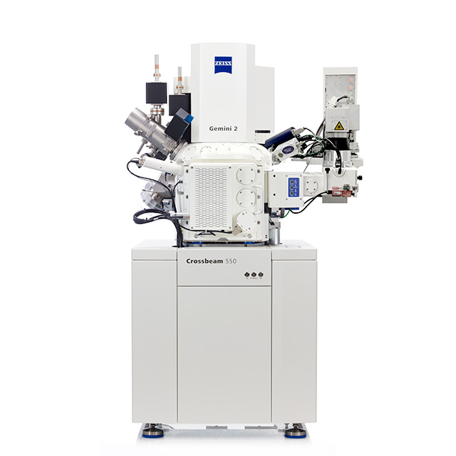
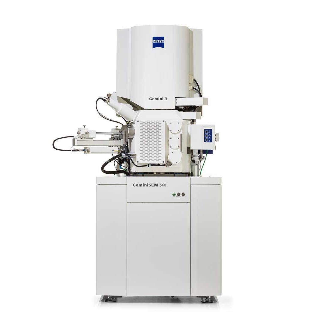
ZEISS Microscopy Solutions for Engineering Materials
Explore our recommended microscopy solutions for engineering materials, designed to meet EPSRC application criteria. This is not an exhaustive list—contact us to discuss your specific needs.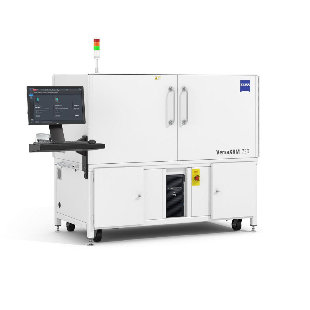


Contact Us
Interested in support for an EPSRC Strategic Infrastructure call application involving a ZEISS solution, or have questions? Complete the form below, and a representative will get in touch.


")
")


")
")