

Microscience Microscopy Congress (MMC) 2025
- 00 years
- 00 months
- 00 days
- 00 hours
- 00 minutes
- 00 seconds
Visit ZEISS at MMC 2025 — Explore the Future of Microscopy
Join us at the Microscience Microscopy Conference (MMC) 2025, where ZEISS will be showcasing the latest in imaging innovation. Stop by Stand 125 to explore a selection of our cutting-edge microscopes in person — including the all-new Lightfield 4D microscope.
You can also book live demo sessions covering both life science and materials science workflows — available on-site and virtually. Spaces are limited, so we encourage early registration.
Plus, don’t miss our two exclusive life science workshops, designed to elevate your insights into imaging even further.
Have Questions? Let’s Chat at Stand 125
Our specialists will be on hand throughout the event to answer your questions and discuss how our newest technologies can support your research. Stop by for a conversation — we’d love to connect.
Demonstration Overview
Get a close-up look at a selection of our systems for life science and materials science — in our focused, expert-led sessions designed to fit your workflow.|
Title |
Description |
Demo Type |
|
|
Life Science Demonstrations |
|||
|
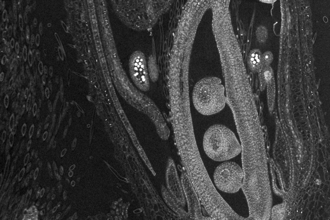
LSM910 with Lightfield 4D |
Be among the first in the UK to experience our NEW LSM with Lightfield 4D, alongside innovations in high-speed volumetric imaging, Airyscan MPLX with jDCV, and our AI-powered microscopy assistant, Copilot. |
In-person Demo |
|
|
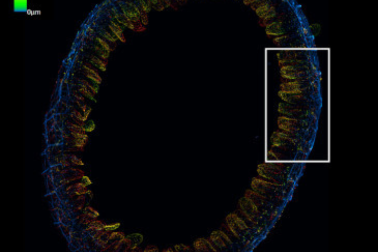
ZEISS Lattice SIM 3 |
Experience fast, high-resolution imaging with ZEISS Lattice SIM 3 — a flexible system that accelerates workflows, captures dynamic processes, and delivers optically sectioned data across a wide range of magnifications. |
In-person Demo |
|
|
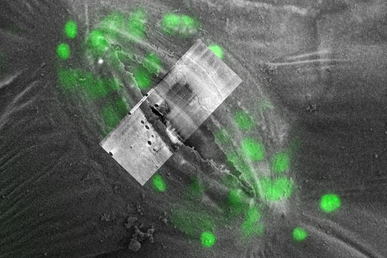
Cryoworkflow - LSM to XB |
Learn how cryo-LM and cryo FIB-SEM combine for targeted, high-resolution 3D imaging of vitrified, immunolabeled samples — from ROI identification to TEM lamella preparation. |
Live Virtual Demo |
|
|
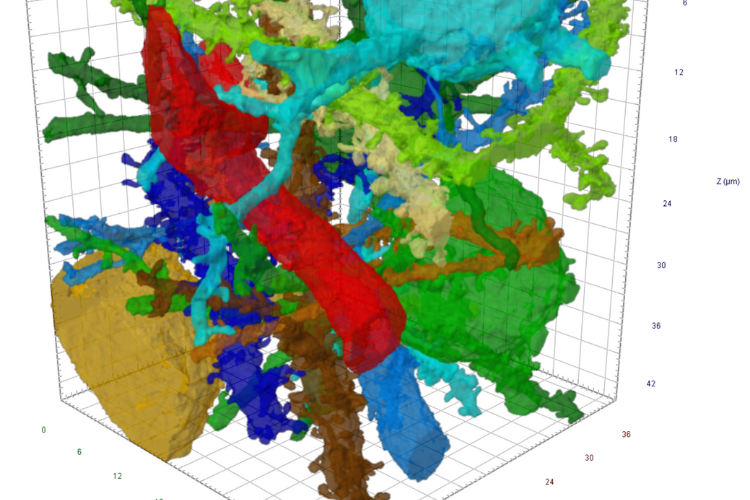
Effortlessly Image Biological Ultrastructure in 3D with ZEISS Volutome |
Explore streamlined 3D ultrastructural imaging with ZEISS Volutome — an integrated SBF-SEM solution for high-quality volume data from resin-embedded samples. |
Live Virtual Demo |
|
|
X-ray Microscopy for Biology |
Discover how X-ray microscopy enables high-resolution 3D imaging of intact biological samples—no sectioning required—using innovations like RaaD and the Advanced Reconstruction Toolbox. |
Live Virtual Demo |
|
|
Materials Science Demonstrations |
|
||
|
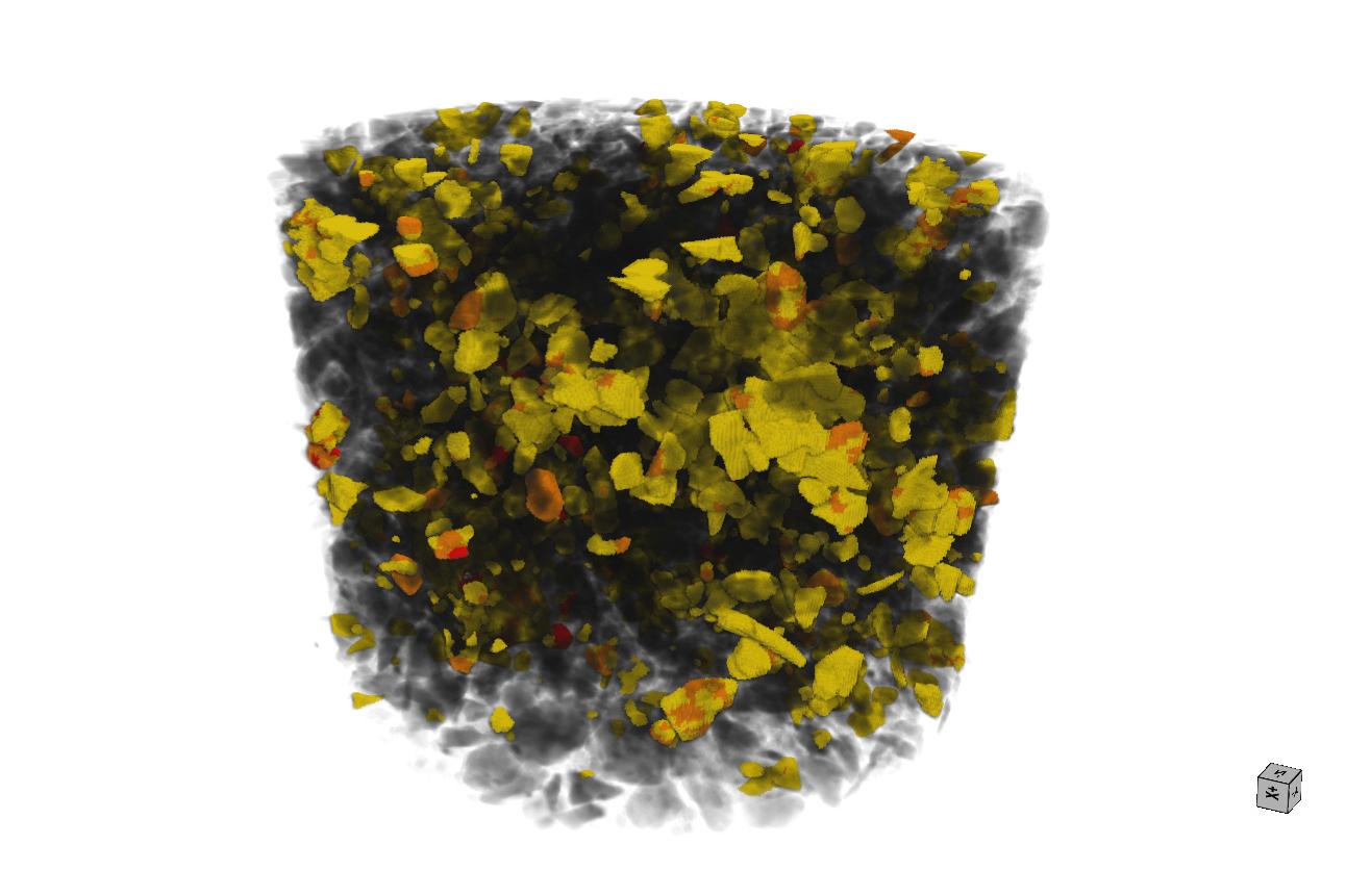
Inside Energy Materials with the ZEISS GeminiSEM 460 |
Register to see how GeminiSEM 460 reveals microstructures in energy materials to support the design and analysis of next-gen storage systems. |
Live Virtual Demo |
|
|
Rapid CT with VersaXRM FAST Mode |
Discover how ZEISS VersaXRM’s FAST Mode delivers rapid, high-quality 3D imaging of complex materials using flat panel technology. |
Live Virtual Demo |
|
|
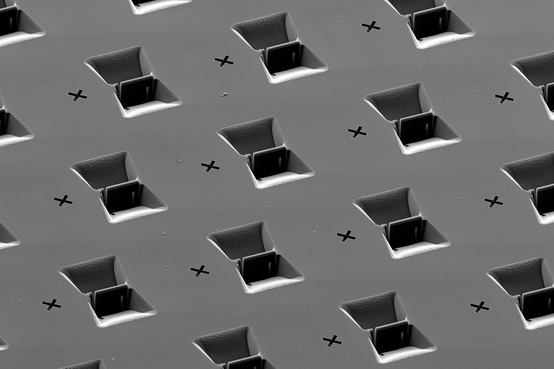
Smart TEM Lamella Preparation with ZEISS Crossbeam |
Experience automated, high-precision TEM lamella prep with ZEISS Crossbeam and ZEN Core for streamlined, high-throughput workflows. |
Live Virtual Demo |
|
|
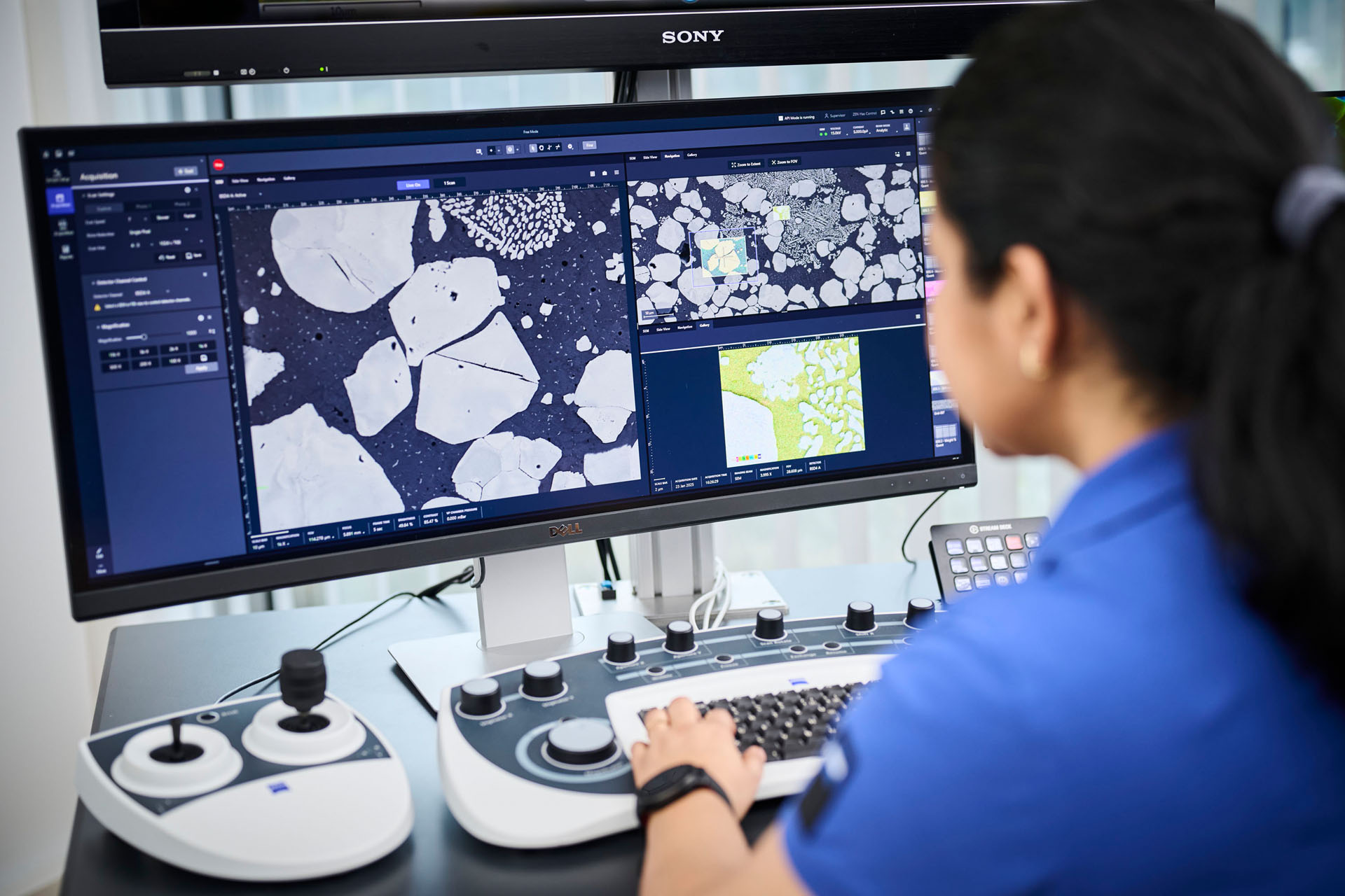
Imaging to Insights with ZEN Core for EM |
Learn how ZEN Core streamlines SEM workflows for faster, more reproducible, and reliable results. |
Live Virtual Demo |
|
|
|
|
|
|
Life Science Demonstrations
Join us for live and virtual demos that reveal new ways to visualize complex biological structures — in full 3D and with minimal disruption. Whether you're working with vitrified samples, resin-embedded tissues, or delicate soft matter, our expert-led sessions will show you how to push the limits of resolution, speed, and sample preservation. Discover how advanced tools can accelerate and enhance your life science research. Register below to save your spot - spaces are limited!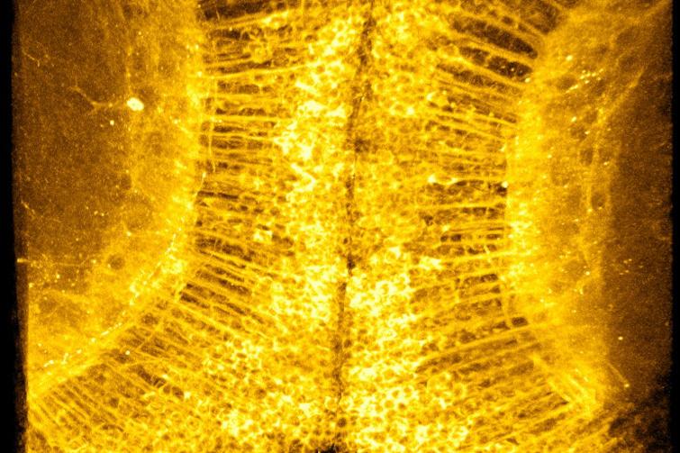


Materials Demonstrations
Join us for live demonstrations—virtual or in person—designed for materials scientists ready to push boundaries. Explore powerful workflows and breakthrough imaging technologies, from ultrafast 3D CT to automated TEM prep and advanced SEM analysis. Whether you're investigating energy materials or streamlining your lab’s throughput, these demos offer hands-on strategies to accelerate your research. Secure your spot today and see the difference firsthand. Register below to save your spot - spaces are limited!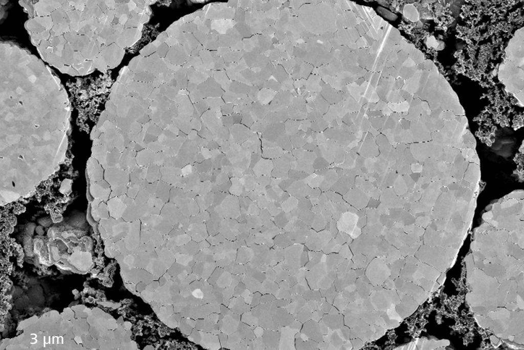


Life Science Workshops
Unlock new insights into dynamic biological processes at ZEISS workshops during MMC 2025. Learn how advanced techniques like Lattice SIM and 4D Light Field Microscopy can elevate your imaging — from faster acquisition to richer spatial detail. Reserve your spot today — limited seats available!
-
Lattice SIM 3 Workshop
Imaging speed is key consideration, whether you’re trying to acquire large areas or fast dynamics and confocal system are typically speed limited. In workshop we will be presenting the Lattice SIM 3, a fast optical sectioning system that utilises structured illumination to give fast, high resolution images. The Lattice SIM mode doubles the resolution of your imaging while allowing collection deep into samples and the SIM Apotome mode gives flexibility across samples sizes to collect large areas or fast dynamics.
-
ZEISS LSM 910 with Lightfield 4D workshop with On stand Workshop
ZEISS are proud to present the UK debut of our new LSM with Lightfield 4D. In this workshop we will focus on a number of new innovations, the first being the new Lightfield 4D with its ability to capture volumes in a single snap and collect volumes at speeds of 80 volumes per second. Continuing the theme of speed, we’ll also explore the new jDCV for Airyscan MPLX. Finally, we will have a discussion with our new AI assistant, Copilot, a powerful LLM based assistant who can aid you in all manner of microscopy challenges.