
Cutting-Edge Solutions for Biomaterials Research
Enhance Your EPSRC Strategic Infrastructure Funding ApplicationAdvance biomaterials research with innovative microscopy solutions- tailored to your needs. Let’s discuss how we can support your application.
Improving human health is one of the greatest challenges of the 21st century, and biomaterials are key to enhancing both lifespan and quality of life. Any material designed for use in the human body requires a deep, precise understanding of its properties to ensure safety and performance. ZEISS microscopy solutions provide cutting-edge imaging techniques for non-conductive materials, along with high-resolution, non-destructive 3D imaging—making them essential tools to leverage for the development and analysis of next-generation biomaterials. Check out our solutions and contact us to explore how we can support your application.
Case Studies: Advancing Research with ZEISS Microscopy
Discover how our cutting-edge microscopy solutions are driving innovation in biomaterials research.

Low kV Imaging
Low kilovolt (kV) operation in Scanning Electron Microscopy (SEM) is essential for analysing sensitive materials and fine features, as it minimises electron beam penetration depth, reducing damage to delicate samples like biological tissues and nanostructures. This is particularly important in semiconductor research for characterising thin films and surface features.
Low kV also enhances surface sensitivity, improving imaging of surface topography while minimising charging effects that can distort signals. Recent developments in low and ultra-low voltage imaging using ZEISS Gemini have demonstrated successful imaging of various materials, including polymers and biomaterials, at voltages between 2 kV and 500 V, allowing for high-resolution imaging of beam-sensitive, non-conductive samples such as polymer battery separators, graphite and chitin.


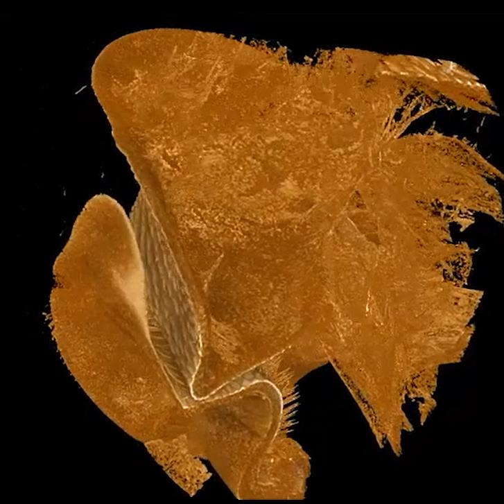
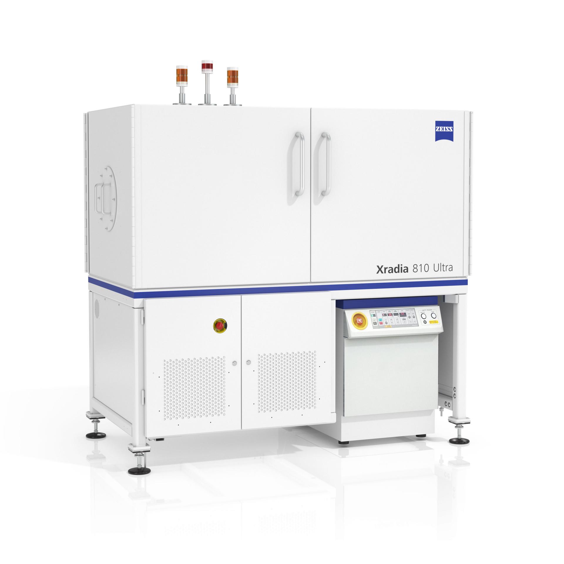
Nano X-ray Microscopy of Electrospun Nanofibre Scaffolds
Tissue engineering focuses on repairing and regenerating damaged organs or tissues using ex vivo created constructs, with a primary challenge being the design of scaffolds that support three-dimensional (3D) tissue growth and wound healing for clinical applications, such as bioartificial transplants. These scaffolds must be biocompatible, highly porous, and possess adequate pore sizes to facilitate nutrient and oxygen diffusion, while also being biodegradable to allow for absorption by surrounding tissues without surgical removal.
Additionally, they should mimic the native tissue's form, architecture, and mechanical stability. The characterisation of scaffold morphology, including porosity and fiber morphology, is essential for optimising materials for specific applications. This study showed the benefits of X-ray microscopy (XRM) characterisation applied to cross-linked electrospun gelatin nanofibers. The unique phase-contrast capabilities of the ZEISS Xradia 810 Ultra microscope were utilised to enhance the contrast in the low density gelatin nanofibers. This allowed for analysis of the effects of fiber cross-linking on gelatin mat morphology, paving the way for improvements in material and process.
ZEISS Microscopy Solutions for Biomaterials
Explore our recommended microscopy solutions for biomaterials, designed to meet EPSRC application criteria. This is not an exhaustive list—contact us to discuss your specific needs.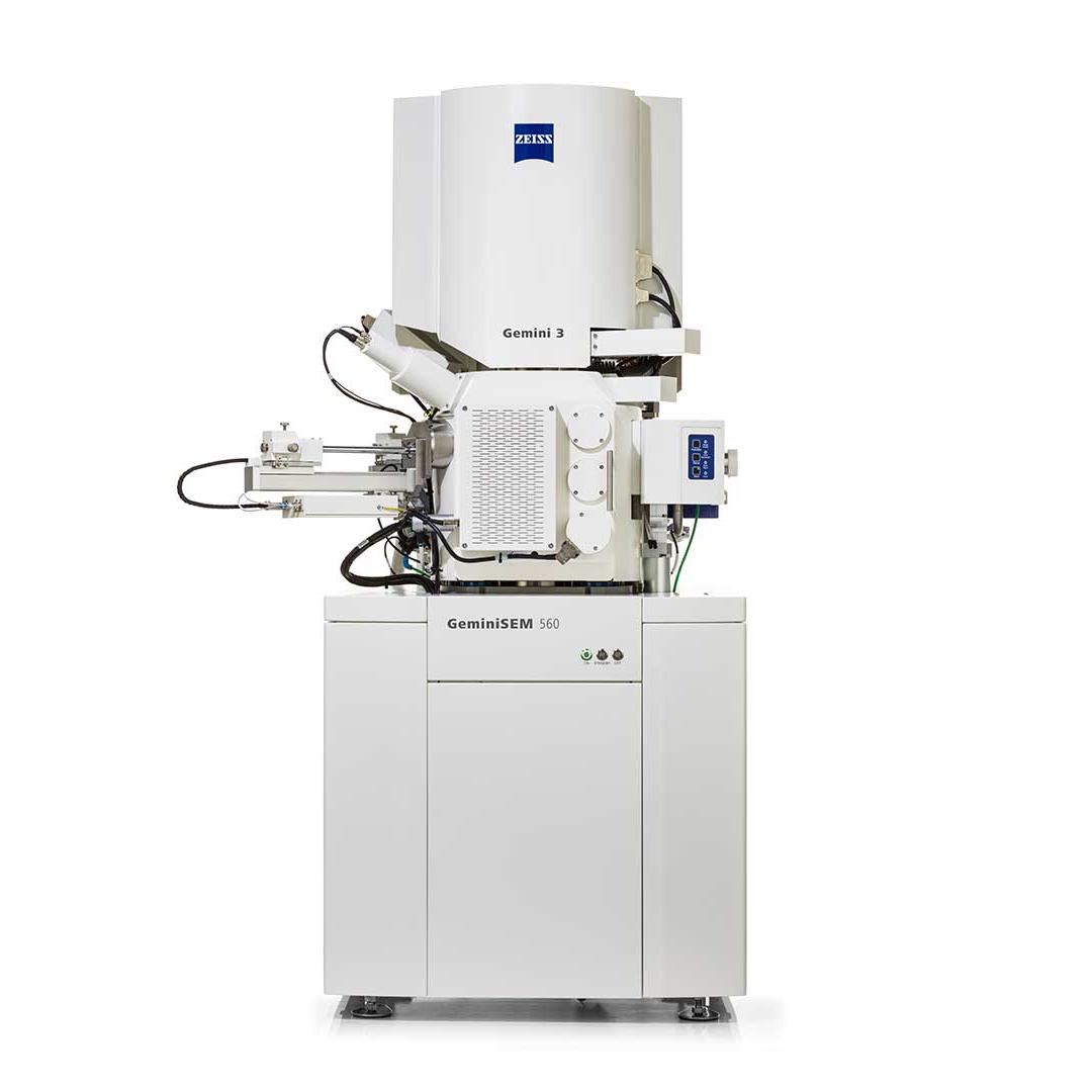


Contact Us
Interested in support for an EPSRC Strategic Infrastructure call application involving a ZEISS solution, or have questions? Complete the form below, and a representative will get in touch.


")
")


")
")


