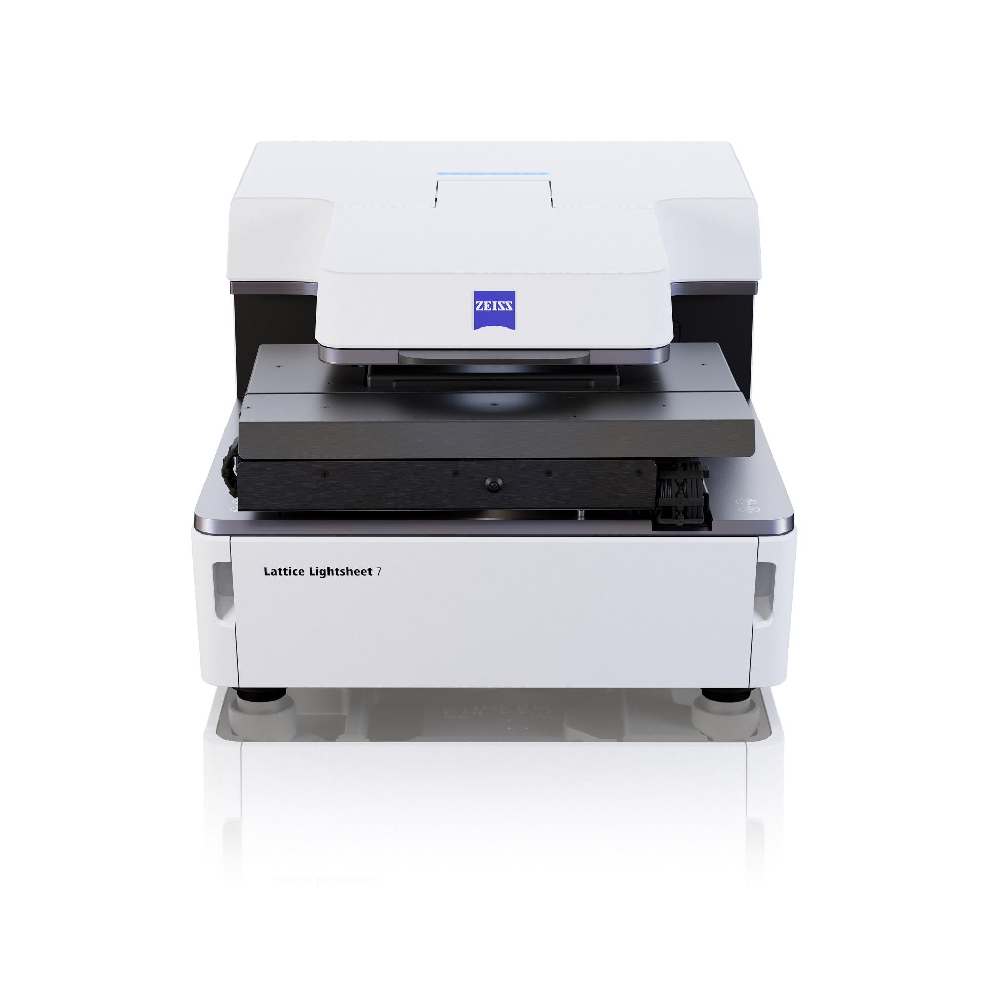

ZEISS Lattice Lightsheet 7
Long-term volumetric imaging of living cellsThe ZEISS Lattice Lightsheet 7 couples cutting edge live cell imaging capabilities with the ease of use you expect from ZEISS. Redefining imaging speed to volumes per second and offering unrivalled gentleness for your live cell experiments. The volumetric imaging at subcellular resolution allows exquisite exploration of dynamics in live samples. The system has been specially designed to work with a range of standard sample carriers, such as slides, dishes and chamber slides, letting you focus on the science, instead of the sample preparation.
Discover the Subcellular Dynamics of Life

Lattice Light Sheet Technology Made Accessible to Everyone
Watch our demonstration in under 60 secondsThe automatic alignments and easy workflows provided by the inverted Lattice Lightsheet 7 instrument mean that every user can now access this cutting-edge approach and capture 3D data of their classically mounted samples over hours and days at a time. Click the video to see the Lattice Lightsheet 7 demonstrated in less than 60 seconds.
Typical Applications

-
- Short-term or long-term observation of physiological and morphological parameters in 2D/ 3D during growth, differentiation, motility and interaction.
- Acquire high resolution images of multi-labelled cell culture from multiwell plates quickly.
- Study the motility of vesicles, organelles and subcellular structures
- Examine the interplay of multiple proteins
- Image subcellular structures at physiological expression levels
- Study molecular dynamic with FRAP
- Explore the interaction of two proteins with FRET
- Observation of stimulus-induced responses of cells or organisms without disturbing the environmental control.
- Perform label free growth curve assays over several days

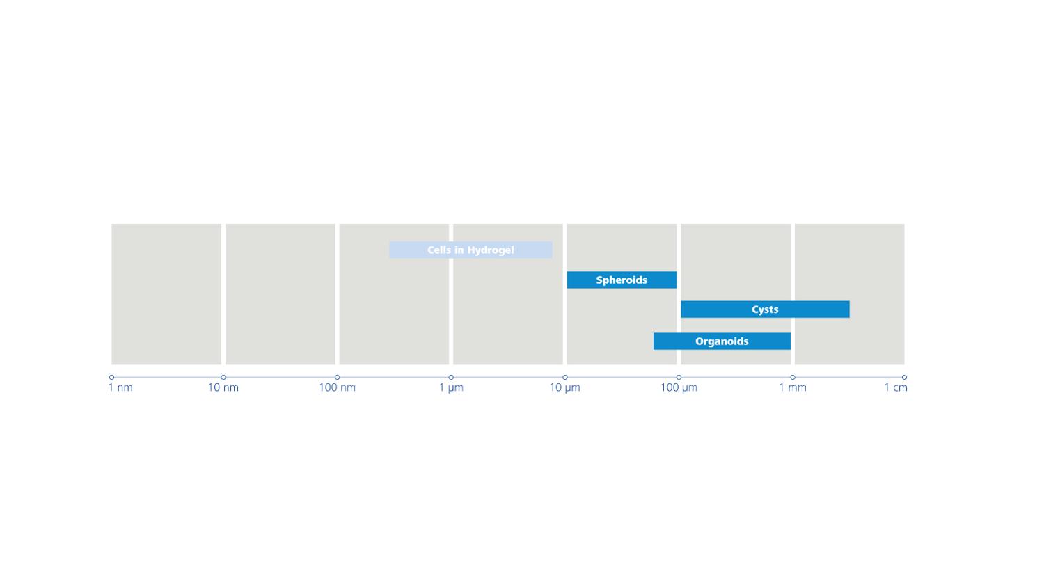
-
- Live imaging of spheroids or organoids with diameters up to 200 μm
- Organoid self-organization
- Cell migration and proliferation within organoids
- Imaging of cell-cell interactions, 3D organization, migration and morphology
- In vitro imaging of neuronal activity
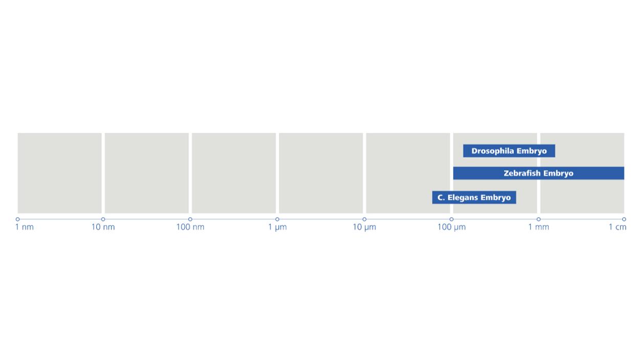
-
- Resolving structural detail in 3D with close to isotropic resolution
- Fast imaging of cellular and subcellular dynamics in embryos and small organisms up to 100 μm in diameter
- Cell migration, cell-cell interaction, cell cycle, vesicle trafficking
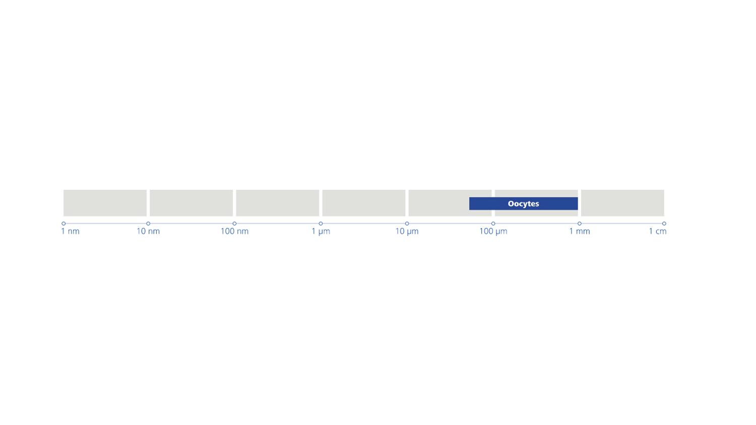
-
- Live imaging of whole oocytes in 3D with subcellular detail


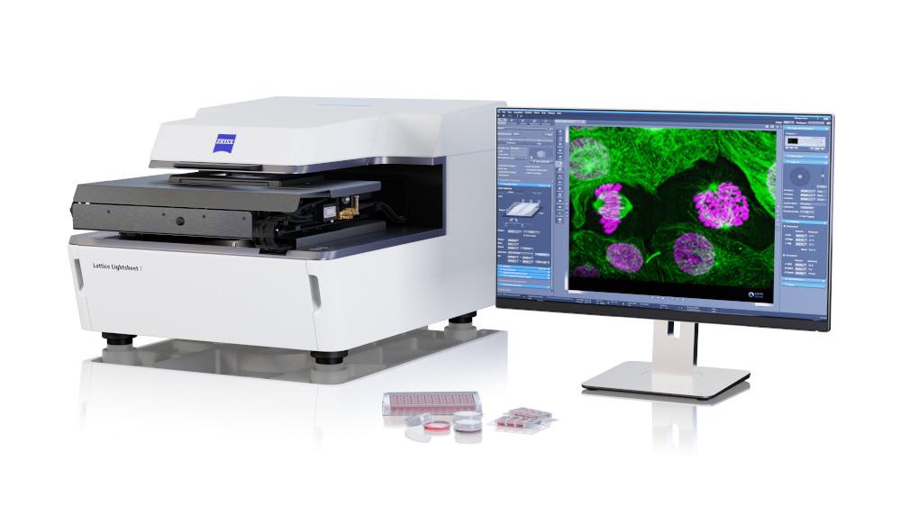
From Image to Results
Image analysis? No ProblemsThe Lattice Lightsheet 7 delivers ease of use for your cutting edge scientific questions, but what about the data? With arivis you can explore and analyse the data rich images from the Lattice Lightsheet 7 in a streamlined way. arivis Vision4D enables users to define and optimise complex image analysis pipelines using easy to navigate tools increasing the efficiency of your research. Data proficiency at everyone’s fingertips.
Trafficking mRNA molecules were tracked in arivis Vision4D®. The movement of the zebrafish embryo was first corrected using a nucleus reference track. Then individual mRNA molecules were tracked over time to result statistics such as speed and directionality. Sample: courtesy of Prof. Andrew Oates, EPFL, Switzerland.
In our new series “From Image to Results” explore how data captured with the ZEISS Lattice Lightsheet 7 can be combined with a powerful Image Analysis Pipeline delivered by arivis Vision4D to generate a high-resolution longitudinal study of vesicle trafficking in Cos7 cells.


