Fuel Cells: Enabling Low Emission Transportation
Explore this Solid Oxide Fuel Cell Anode, scanned non-destructively in 3D with Nanoscale ZEISS X-ray Microscopy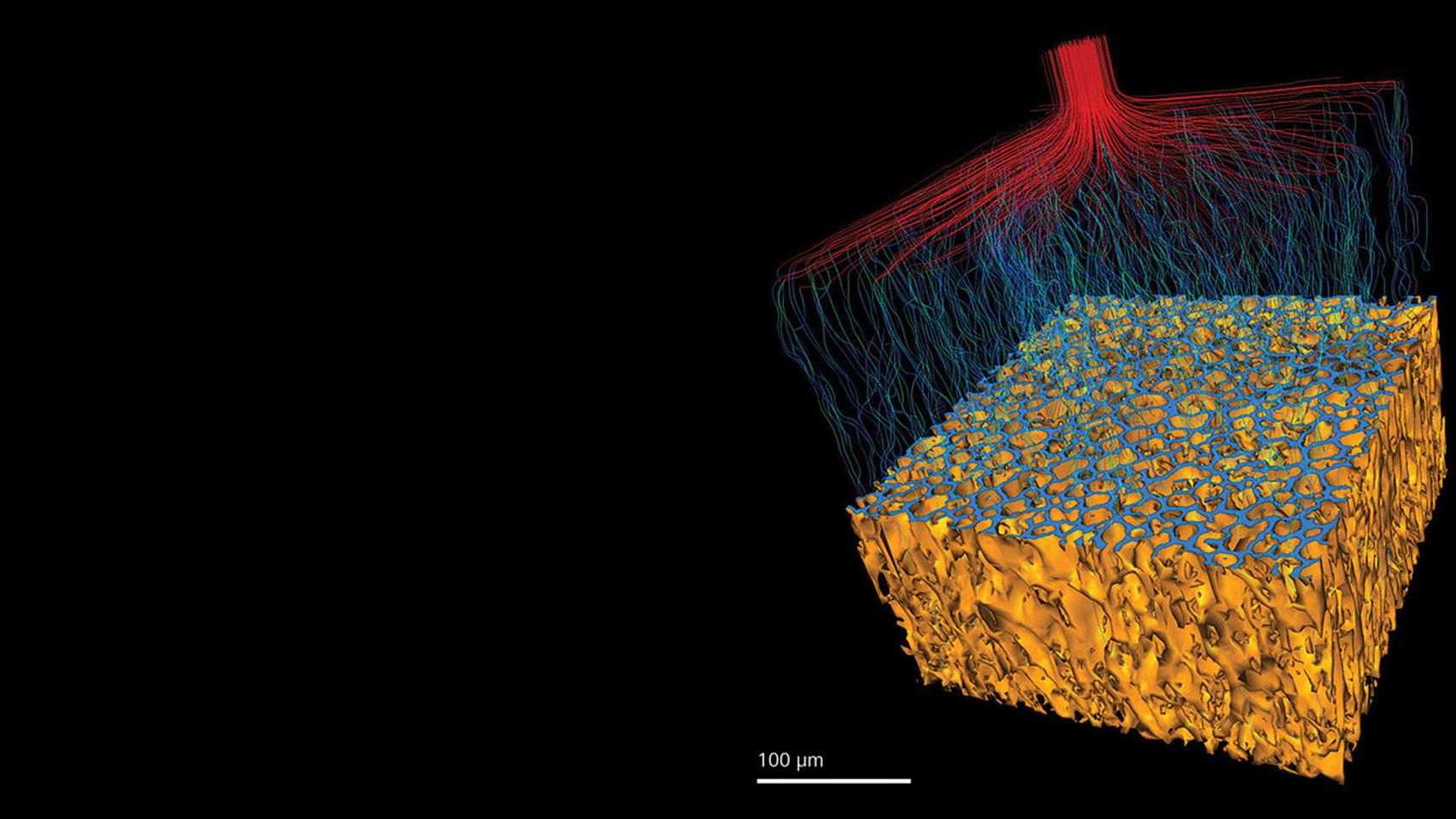

As mentioned in the above video, this dataset is an anode of a solid oxide fuel cell used in the conversion of hydrogen to electricity. The sample was scanned non-destructively in 3D using X-ray Microscopy with the ZEISS Xradia Ultra.
3D deep-dive into the sample with X-ray
Microscopy Sample courtesy of Sandrine Ricote, Colorado School of Mines.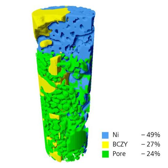

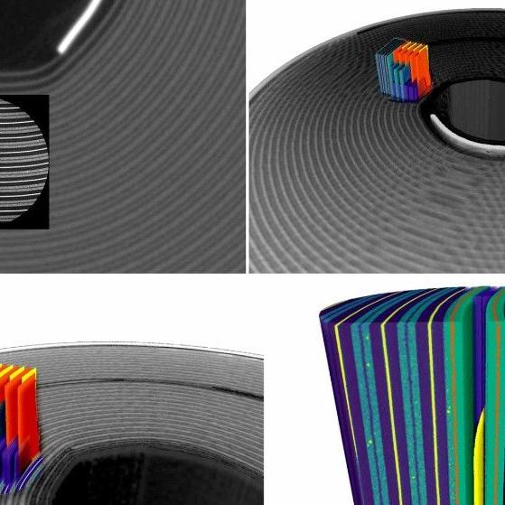
Figure 1 shows the large field of view scan. There are three distinct phases identified – metallic nickel, yttria-stabilised zirconia and porosity.
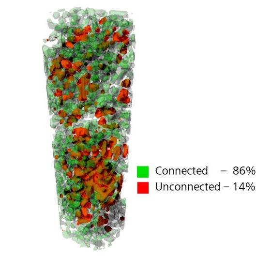

Figure 2 shows a connectivity study, with the majority of pores connected. Connectivity is important as reactants must be able to diffuse for reactions to occur. With pores connected diffusion is easier.
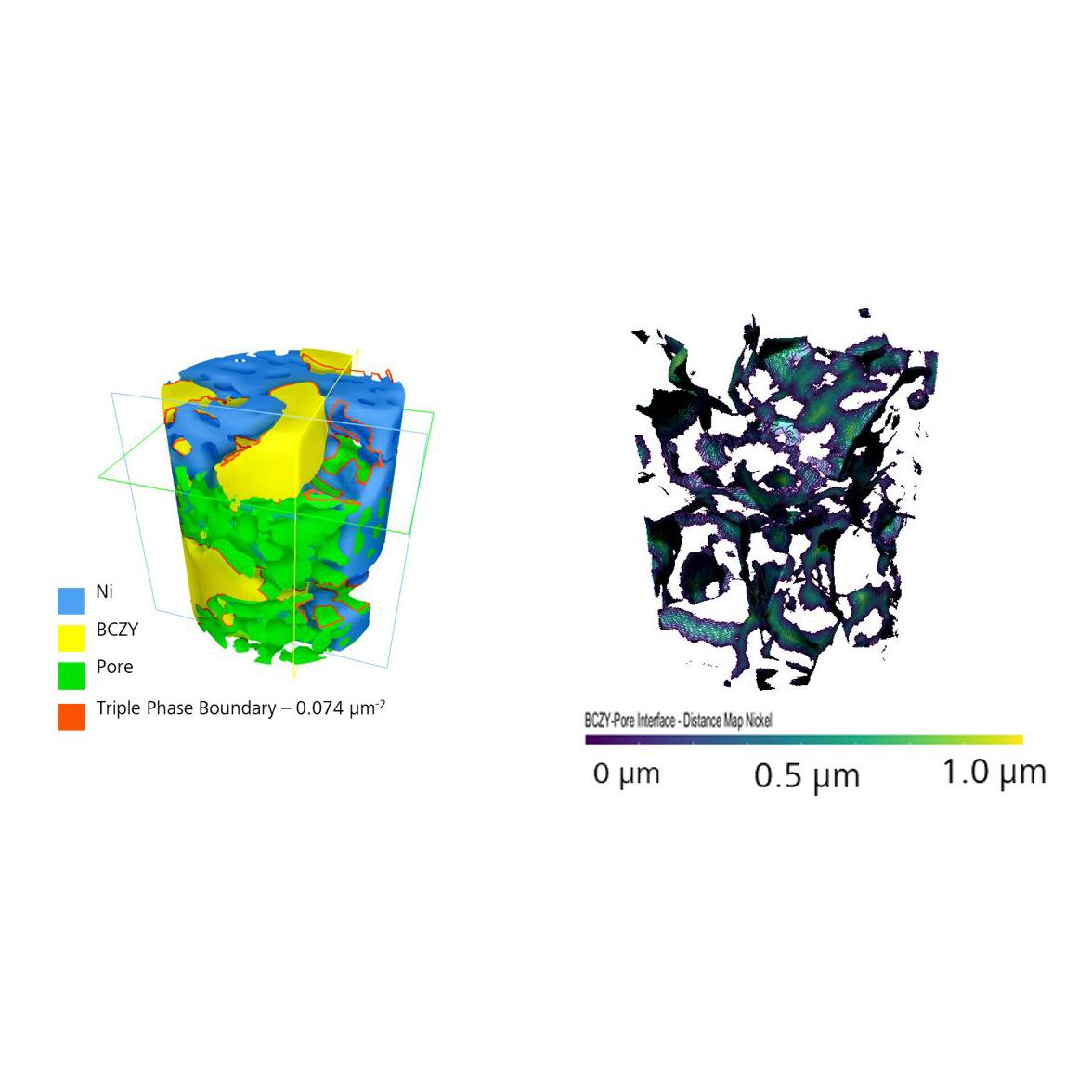

In Figure 3, a high-resolution field of view has been used, increasing resolution the expense of field of view. With the added resolution it is possible to map triple phase boundaries, seen in Figure 4. Triple phase boundaries are the surface areas where reactions can occur, affecting the electrochemical performance of the cell.

ZEISS Xradia Ultra Family
Nanoscale X-ray Imaging – Explore at the Speed of Science3D & 4D nanoscale X-ray imaging with synchrotron-adapted optics and in situ extensions with Xradia 800 & 810 Ultra.
3D in 3 Minutes
See the scanning technology in actionOn-Demand Webinars

Microscopy for Battery Research - An Introduction
Webinar Jun 11, 2020Stephen T. Kelly, Ph.D Solutions Manager, ZEISS X-ray Microscopy


Nanoscale 3D X-ray Imaging for the Laboratory with ZEISS Xradia Ultra
Webinar Jun 16, 2020Dr. Mohsen Samadi, Business Development Manager X-ray Microscopy Systems...

In Situ Lab- and Synchrotron-based X-ray Microscopy Applied to...
Webinar Feb 18, 2021Dr. Johanna Nelson Weker / Geoff McConohy, Stanford Synchrotron Radiation...

