Composites: The Future of Transportation
Explore this Ceramic Matrix Composite, scanned non-destructively in 3D with ZEISS X-ray Microscopy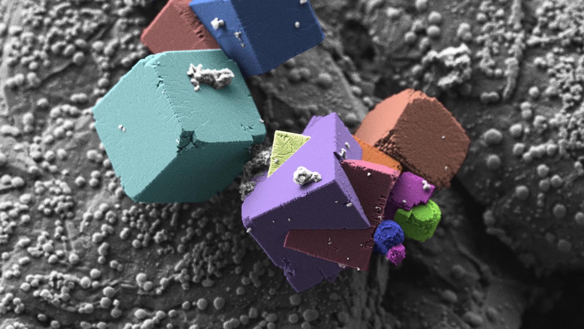

As mentioned in the video above, this dataset is a woven composite comprising of carbon fibres in a silicon carbide matrix. The SiC matrix has been applied using chemical vapor infiltration. The sample was scanned non-destructively in 3D using X-ray Microscopy with the ZEISS Xradia Versa. The Scout & Zoom workflow was used to capture data across length scales from the whole sample overview to the highest resolution scan, where it is possible to detect the fibre clusters.
3D deep-dive into the sample with X-ray
Microscopy Sample courtesy of Sandrine Ricote, Colorado School of Mines.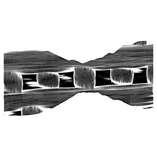

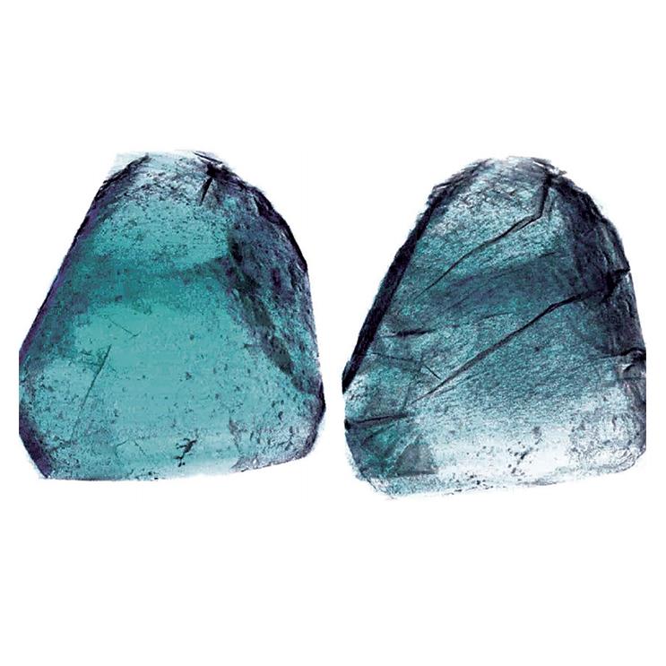
Figure 1 shows the sample overview scanned with the 0.4x objective. Already three distinct phases are visible – C-fibre, SiC and air. Voxel size (a 3D pixel) is 14 microns, so it is not yet possible to see much of the fibre structure.
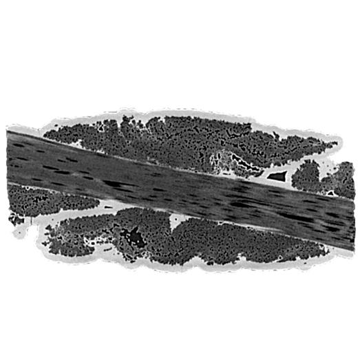

In Figure 2, the 4x objective of the Xradia Versa has been used, giving a higher resolution image at the expense of field of view. There are still 3 phases, but it is now possible to see some detail in the fibre structure. It can be seen that the CVI method used to implant the SiC has not been entirely successful in coating the C-fibres.
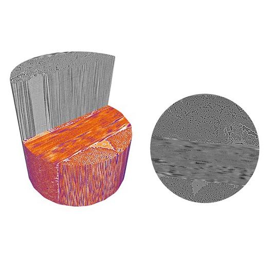

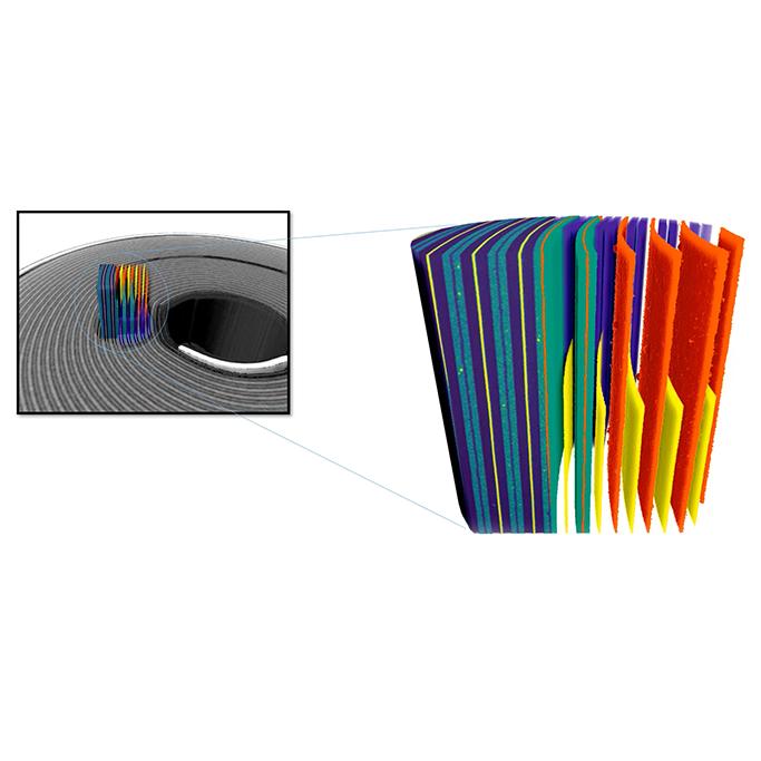
Finally, Figures 3a and 3b show the highest resolution scan, captured using the 20x objective with a voxel size of 0.8 microns. It is now possible to make out individual fibres, along with thin SiC coatings and air voids. With this data an accurate segmentation of the fibre bundles is possible, denoted in orange.
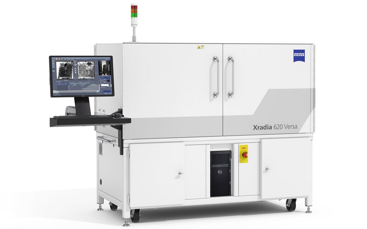

Xradia 610 & 620 Versa
3D X-ray Microscopy for Faster Sub-Micron Imaging of Intact SamplesExpand the boundaries of your non-destructive sub-micron scale imaging built on industry-best resolution and contrast.
3D in 3 Minutes
See the scanning technology in actionOn-Demand Webinars

Opportunities in 3D and 4D Imaging with Laboratory X-ray Microscopy
Webinar Apr 27, 2020Dr. Nicolas Gueninchault, X-Ray Microscopy Systems Product Application and...

In Situ and 4D Characterization Across Length Scales in Materials...
Webinar May 6, 2020Dr. Hrishikesh Bale, ZEISS Microscopy and Dr. Nikhilesh Chawla Fulton...

New Methods for Characterizing Nanomaterials - Exploring Advances in...
Webinar Apr 8, 2020Hrishikesh Bale, Solutions Manager, ZEISS X-ray Microscopy

