ZEISS Celldiscoverer 7 with LSM 900 and Airyscan 2
Your automated microscope for gentle and fast confocal 4D imagingWith Celldiscoverer 7, you combine the ease-of-use of an automated high-end imaging system with the image quality and flexibility of a classic inverted research microscope. Celldiscoverer 7 calibrates itself, detects and focuses on your samples while the optics adjust themselves.
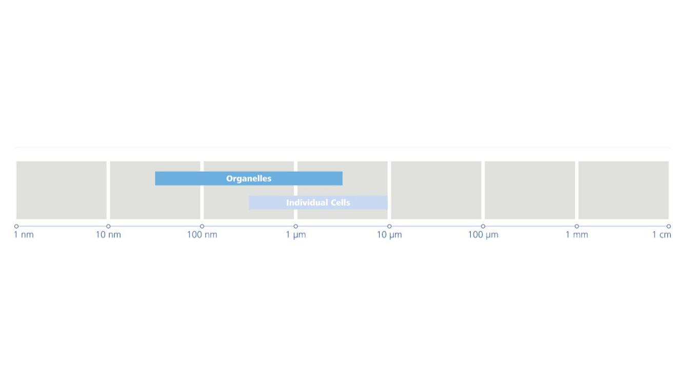
Life sciences research often calls for optical sectioning to image samples with best possible contrast and resolution. The Celldiscoverer 7 with LSM 900 and Airyscan 2 enables gentle 3D imaging of dynamic processes in your living samples with highest framerates in superresolution and easy separation of multiple labels with spectral imaging.
Reproducible Results Made Easy


Case Study: Life with the Celldiscoverer 7
Discover how the Celldiscoverer 7 is changing researchWe spoke with Serge Mostowy, Professor of Cellular Microbiology, and his team at the London School of Hygiene and Tropical Medicine, UK to uncover the transformative impact of the ZEISS Celldiscoverer 7 on their research.

Download Below
Whitepaper:
The transformative impact of the ZEISS Celldiscoverer 7The ZEISS Celldiscoverer 7 was first installed in the Mostowy Lab at the London School of Hygiene & Tropical Medicine in 2019. Since then, the system has enabled them to evolve both their research and imaging related questions - download the White Paper to find out more.
From Image to Results
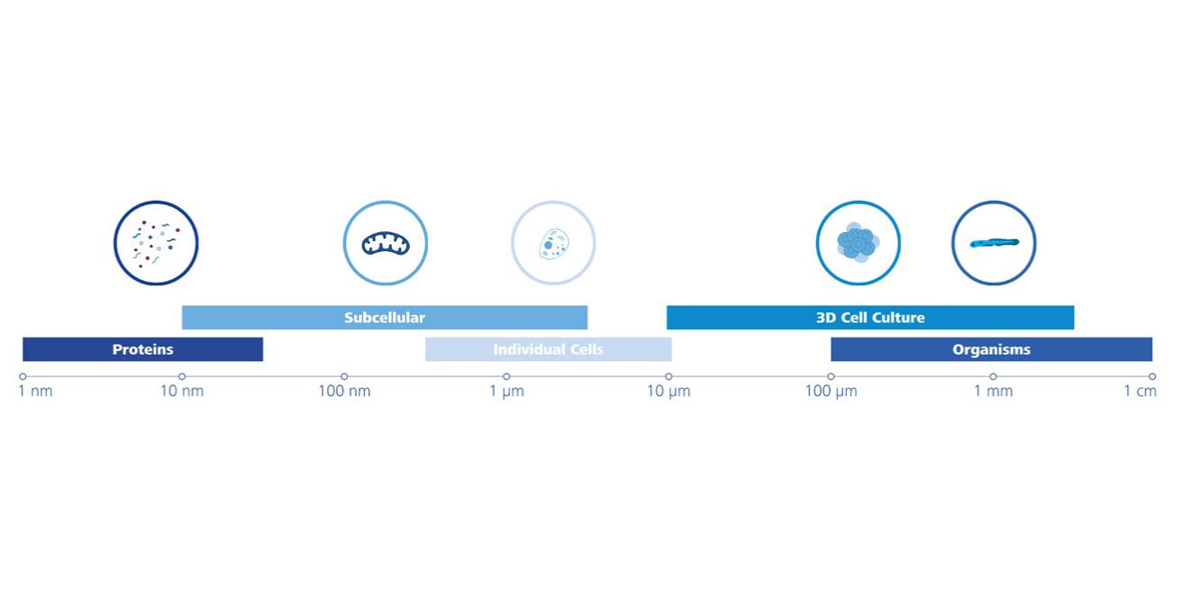
Image analysis? No ProblemsTypical Applications

-
- Short-term or long-term observation of physiological and morphological parameters in 2D/ 3D during growth, differentiation, motility and interaction.
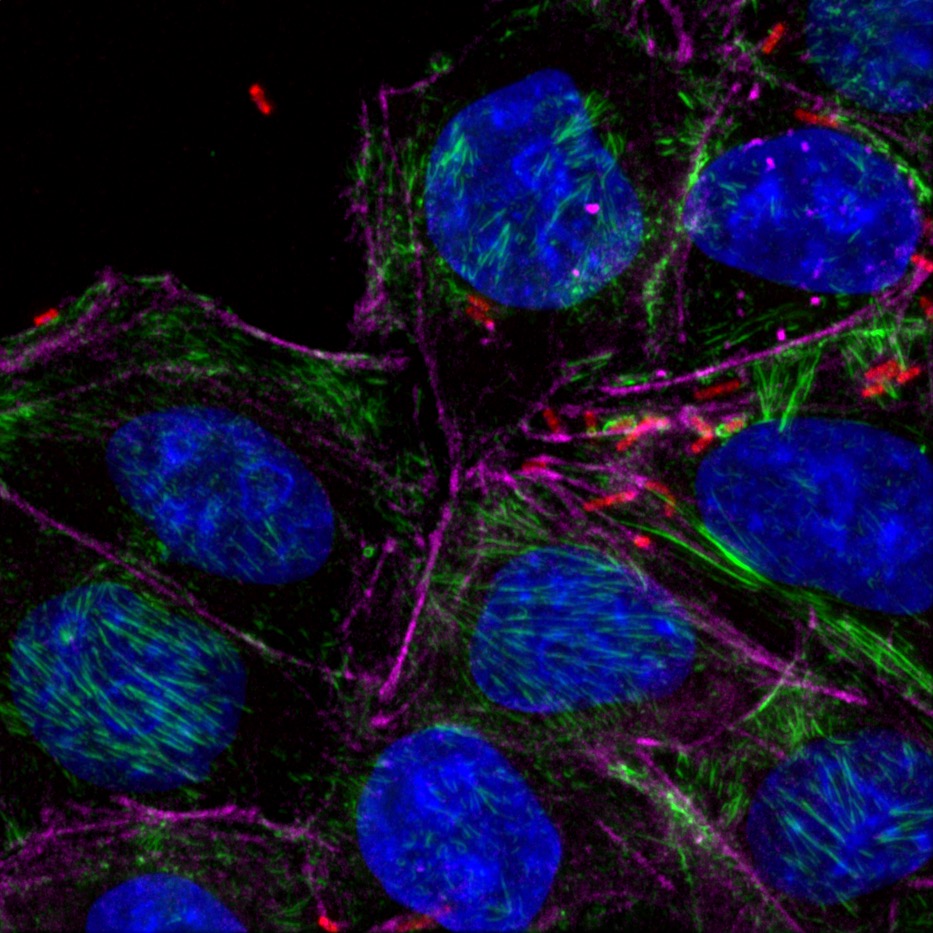
- Acquire high resolution images of multi-labelled cell culture from multiwell plates quickly.
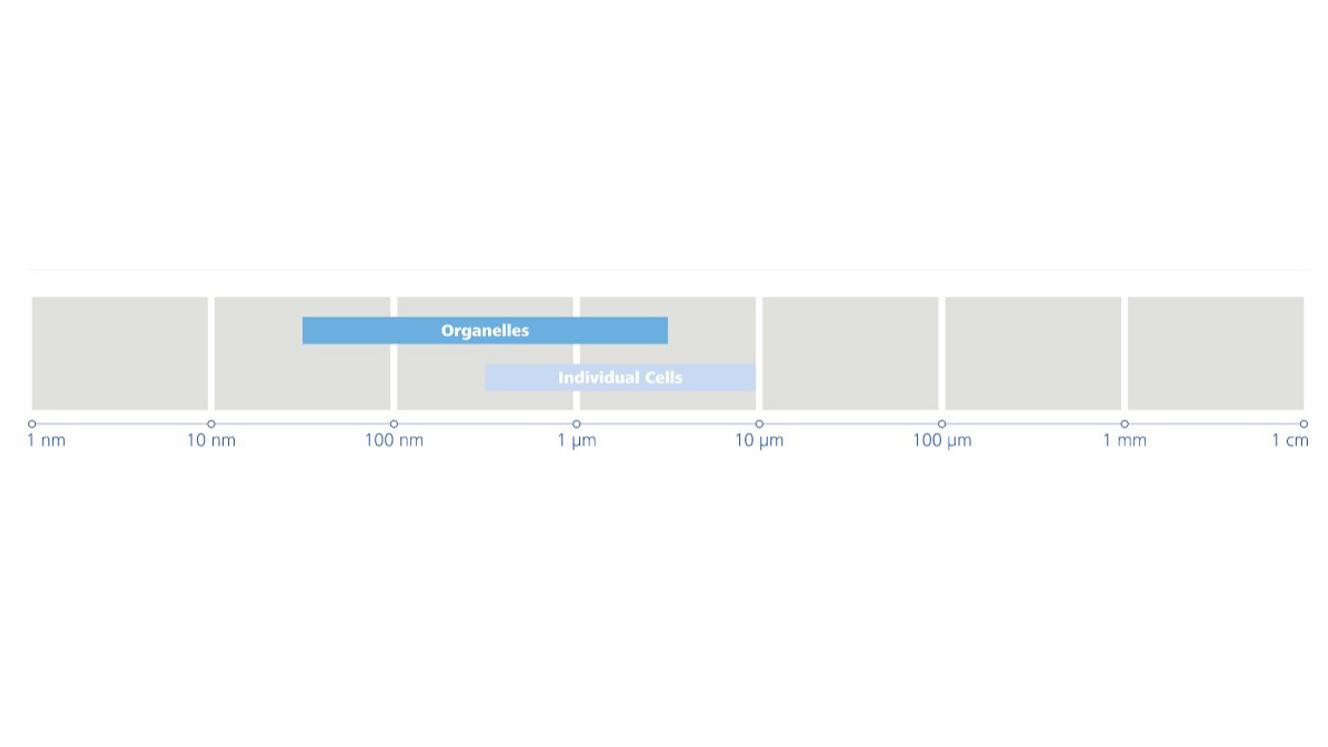
- Study the motility of vesicles, organelles and subcellular structures
- Examine the interplay of multiple proteins
- Image subcellular structures at physiological expression levels
- Study molecular dynamic with FRAP
- Explore the interaction of two proteins with FRET
- Observation of stimulus-induced responses of cells or organisms without disturbing the environmental control.
- Perform label free growth curve assays over several days

-
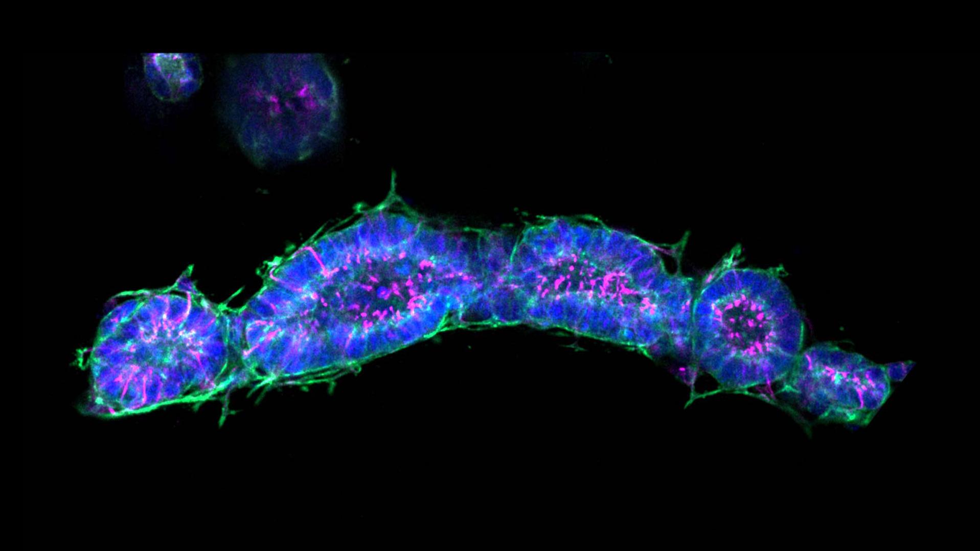
- Live imaging of spheroids or organoids
- Organoid self-organization
- Cell migration and proliferation within organoids
- Study morphogenetic process using tubule organoids.

-
- Cell migration and proliferation within organoids
- Study morphogenetic process using tubule organoids.
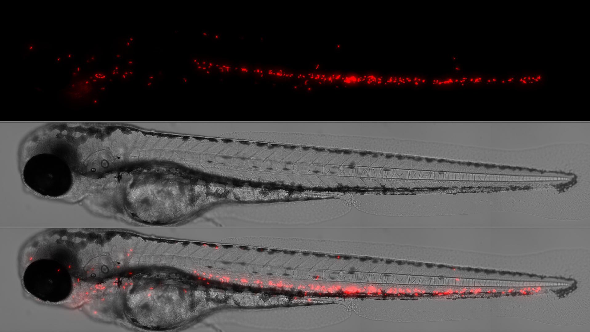
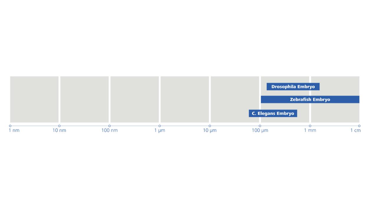
- Analyse the embryogenesis of small model organisms
-
- Change quickly between large overview scans and high resolution imaging
- Fixed fluorescently labelled tissue, cell culture samples - Identification, quantification and qualification of cell types, pathological and pharmacological pathways using cell, tissue and protein markers in 2D and 3D samples.
- Label-free fixed and thin tissue slices or small organisms